
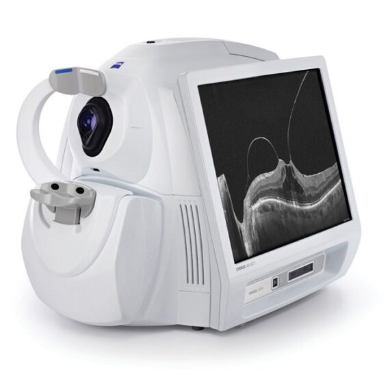

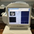
Carl Zeiss Cirrus HD-OCT 5000
$8.950$13.250
CIRRUS HD-OCT 5000 is configured with the highest resolution visualization capabilities and sophisticated clinical applications such as Advanced RPE analysis to track retinal pigment epithelial integrity and Ganglion Cell Analysis to assess glaucomatous loss in the macula that may not be evident in the peripapillary region. The new FastTrac™ retinal tracking reduces eye motion artifacts without sacrificing patient throughput with a proprietary scan acquisition strategy, high speed 20 Hz LSO camera, and single-pass alignment scanning. This feature enables the highest resolution B-Scans to be captured in identical locations from visit to visit providing precise assessment of change in targeted pathologies.
Description
CARL ZEISS Cirrus HD-OCT 5000 provide highest resolution visualization capabilities and track retinal pigment epithelial integrity using Advanced RPE Analysis and assess glaucomatous loss with Ganglion Cell Analysis
Cirrus HD-OCT 5000 come with features an active eye-tracking mechanism that able to trace and compensate eye motion in real time and FastTrac retinal tracking to reduces eye motion artifacts without sacrificing patient
Cirrus OCT come with version 9.5 software can acquire 68,000 scans per second with fundus photos
This Cirrus HD-OCT 5000 completely refurbished by Zeiss trained technician
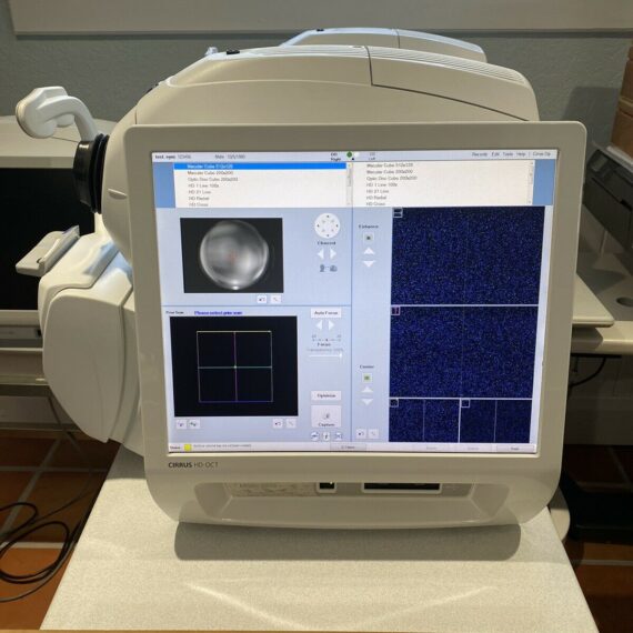
The new updated Cirrus 5000 HD OCT system features state-of-the-art hardware and software based on a fast 64-bit Windows 7 computer, a significantly accelerated OCT camera system, a much larger 19-inch monitor, and a wireless mouse and mouse Keyboard. This results in faster processing with shorter times for the patient and ophthalmologist. The OCT scan speed is between 27,000 and 68,000 per second.
Cirrus 5000 OCT – the big brother
Last but not least, there was an extraordinarily wide range of tools for diagnosing glaucoma, maculopathies, and the anterior segment without having to install an additional lens.
Specifications
- OCT Imaging
- Methodology: Spectral domain OCT
- Optical source: Superluminescent diode (SLD), 840 nm
- Scan speed: 27K- 68K A-scans per second
- A-scan: 2.0 mm (in tissue), 1024
- Axial resolution: 5 μm (in tissue)
- Transverse resolution: 15 μm (in tissue)
Fundus Imaging
- Methodology: Line scanning ophthalmoscope (LSO)
- Live fundus image: During alignment and during OCT scan
- Optical source: Superluminescent diode (SLD), 750 nm
- Field of view: 36 degrees W x 30 degrees H
- Frame rate: > 20 Hz
- Transverse resolution: 25 μm (in tissue)
Iris Imaging
- Methodology: CCD camera
- Resolution: 1280 x 1024
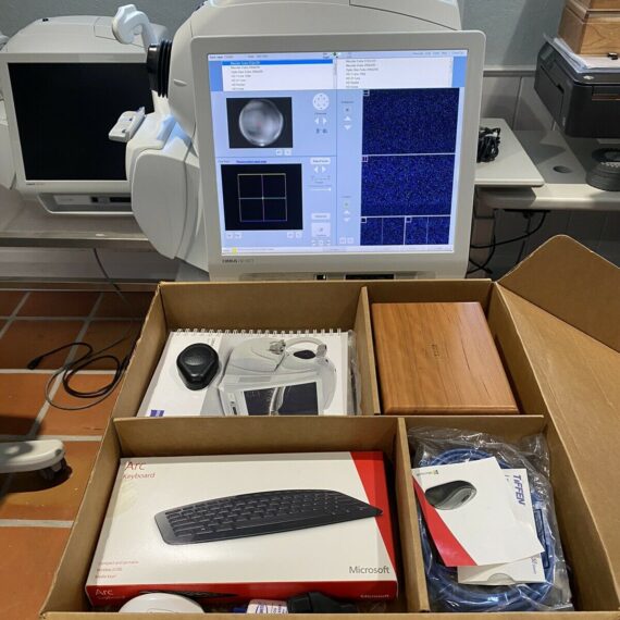
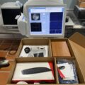
Include
- Cirrus HD-OCT 5000 Unit
- New Printer
- Mouse
- Keyboard
- Warranty Table
- Table
- all peripherals.


Reviews
There are no reviews yet.